Orthognathic Surgery Medical Illustrations
Scroll down to learn about the medical illustrations we created for the Department of Oral and Maxillofacial Surgery.
Client: KU Leuven
01 - Overview
Services:
2D Illustration
Artists:
Margot Ceelen
Ezra van Hattem
Tiffany Fung
We collaborated with the KU Leuven to produce a large series of medical illustrations visualizing the historical surgical methods used in oral and maxillofacial surgery. This series is aimed as teaching material for the surgeons-in-training at the academic hospital.
A Look into the Le Fort Osteotomy
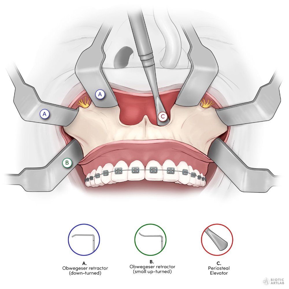
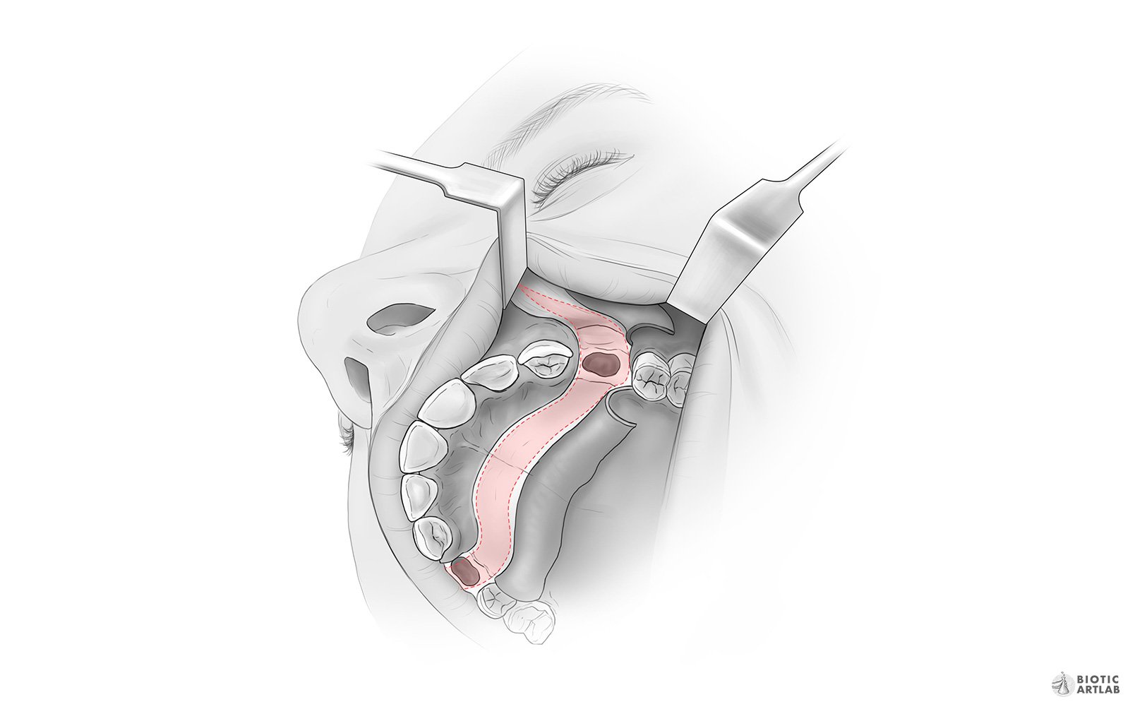
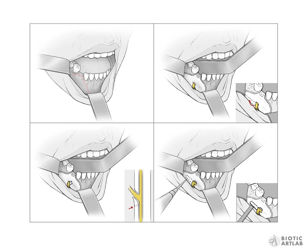
Step 1: An incision is made into the gums to reveal the maxilla.
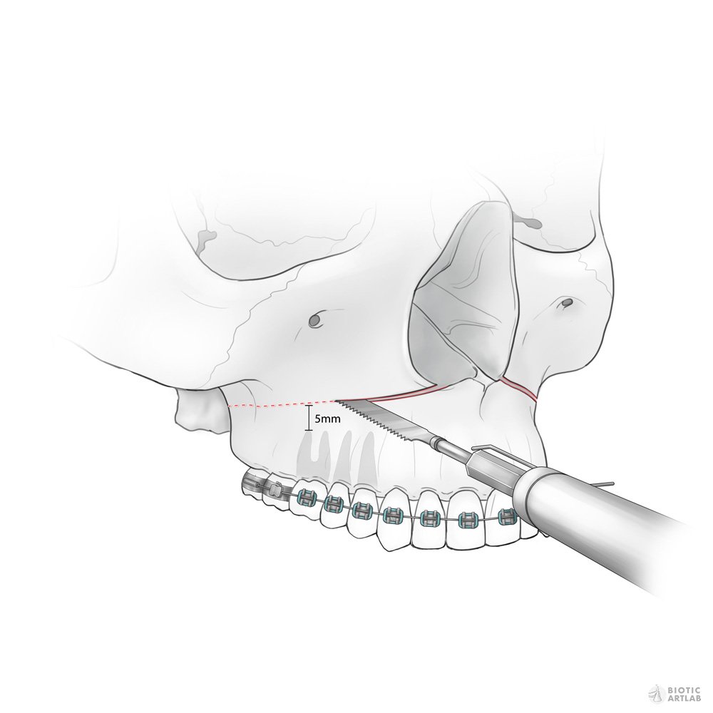
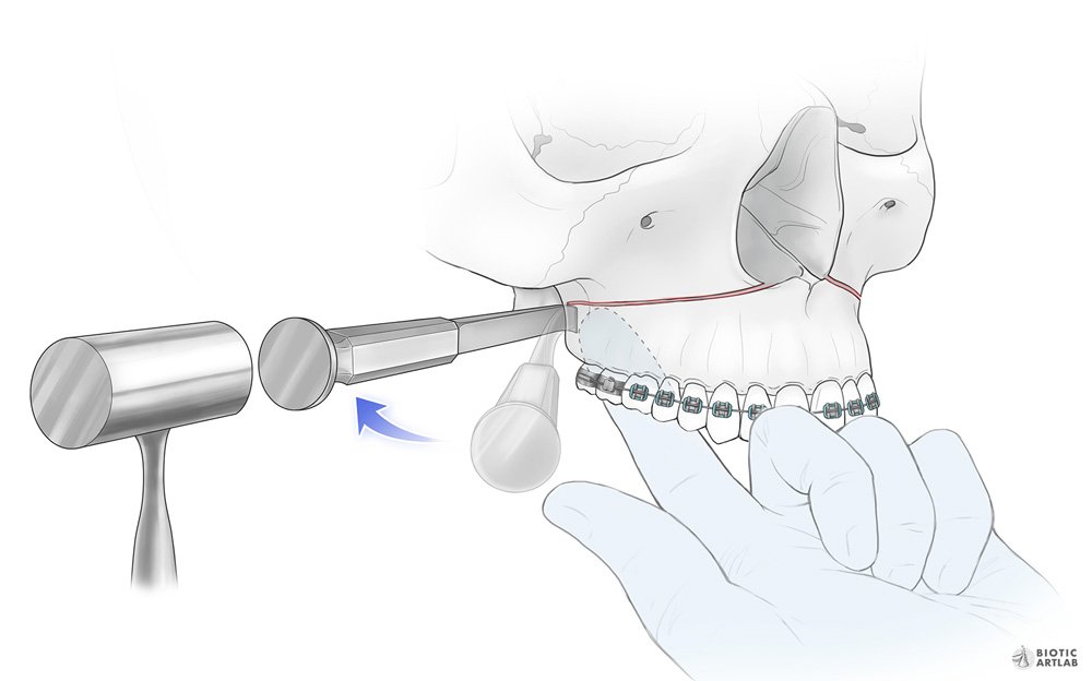
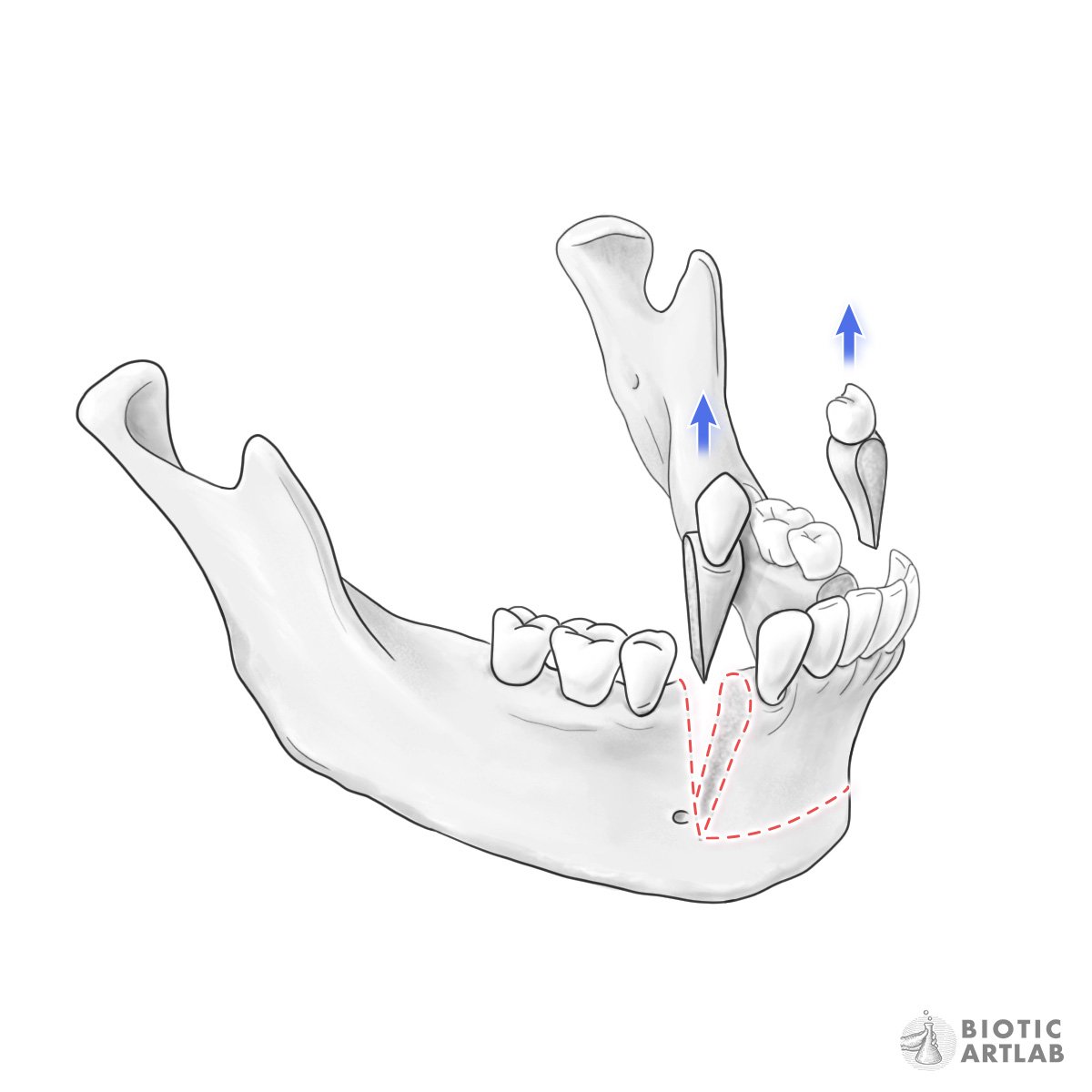
Step 2: A bone saw is used to create the osteotomy from nasal cavity to pterygoid junction.
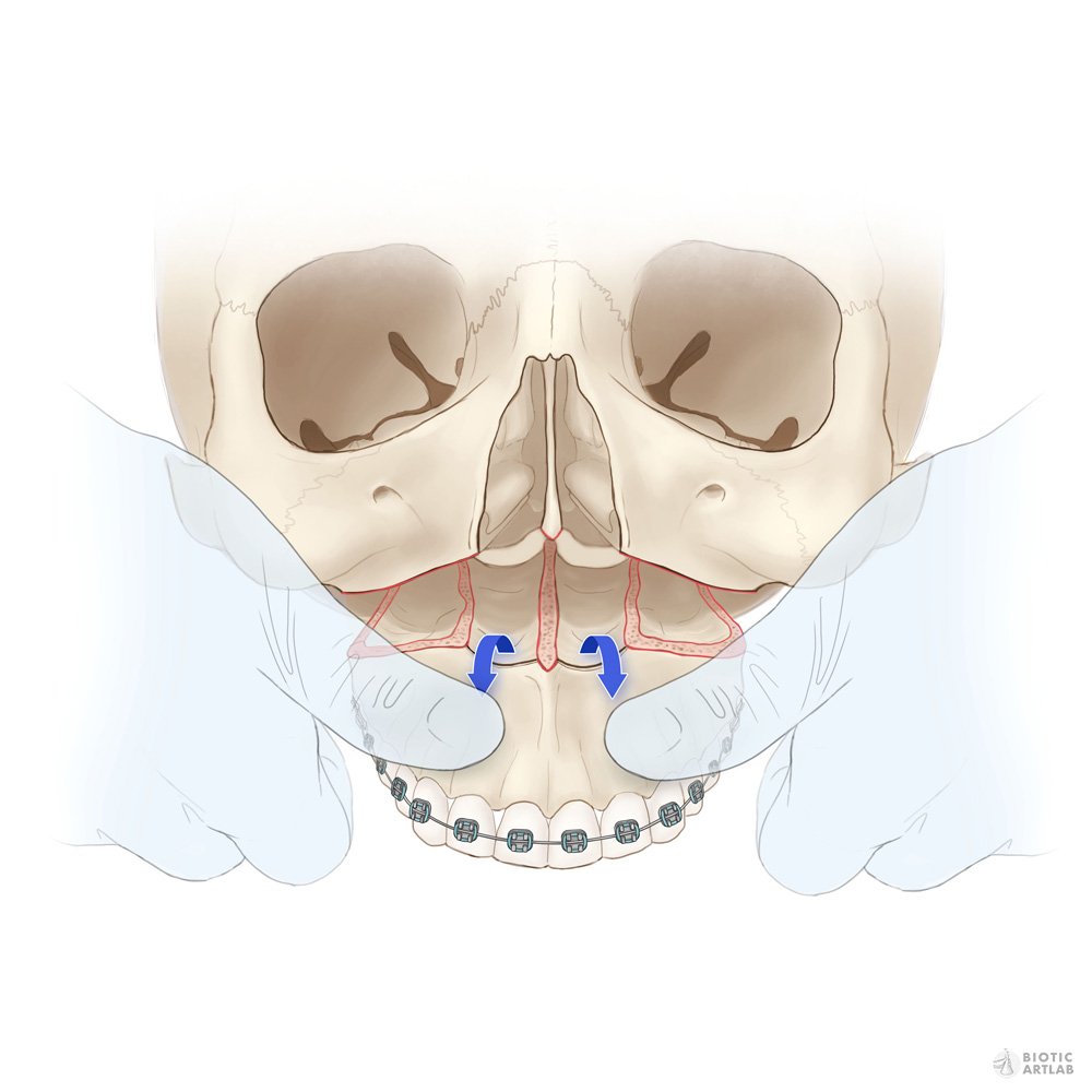
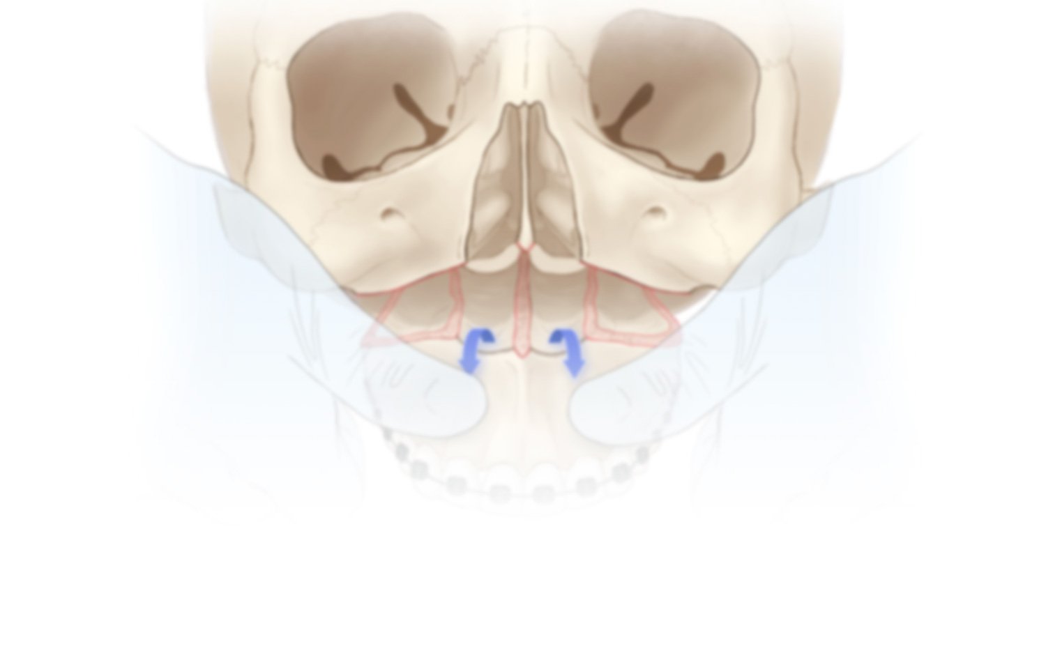
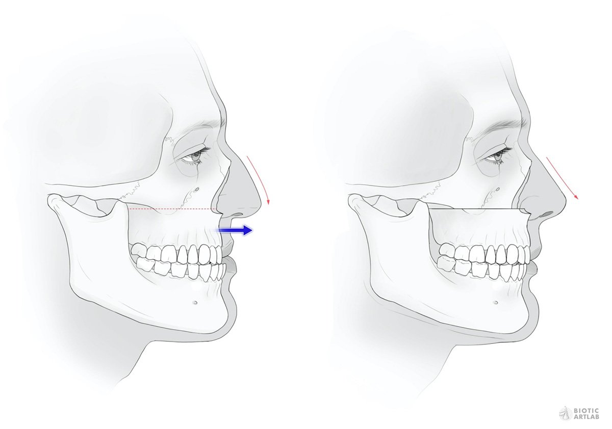
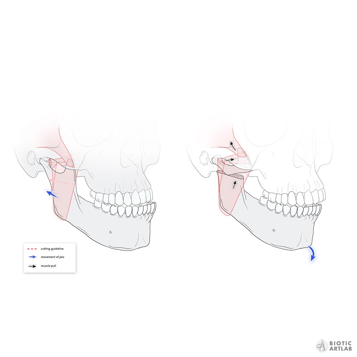
Step 3: Manual pressure is used to break the maxilla downwards.
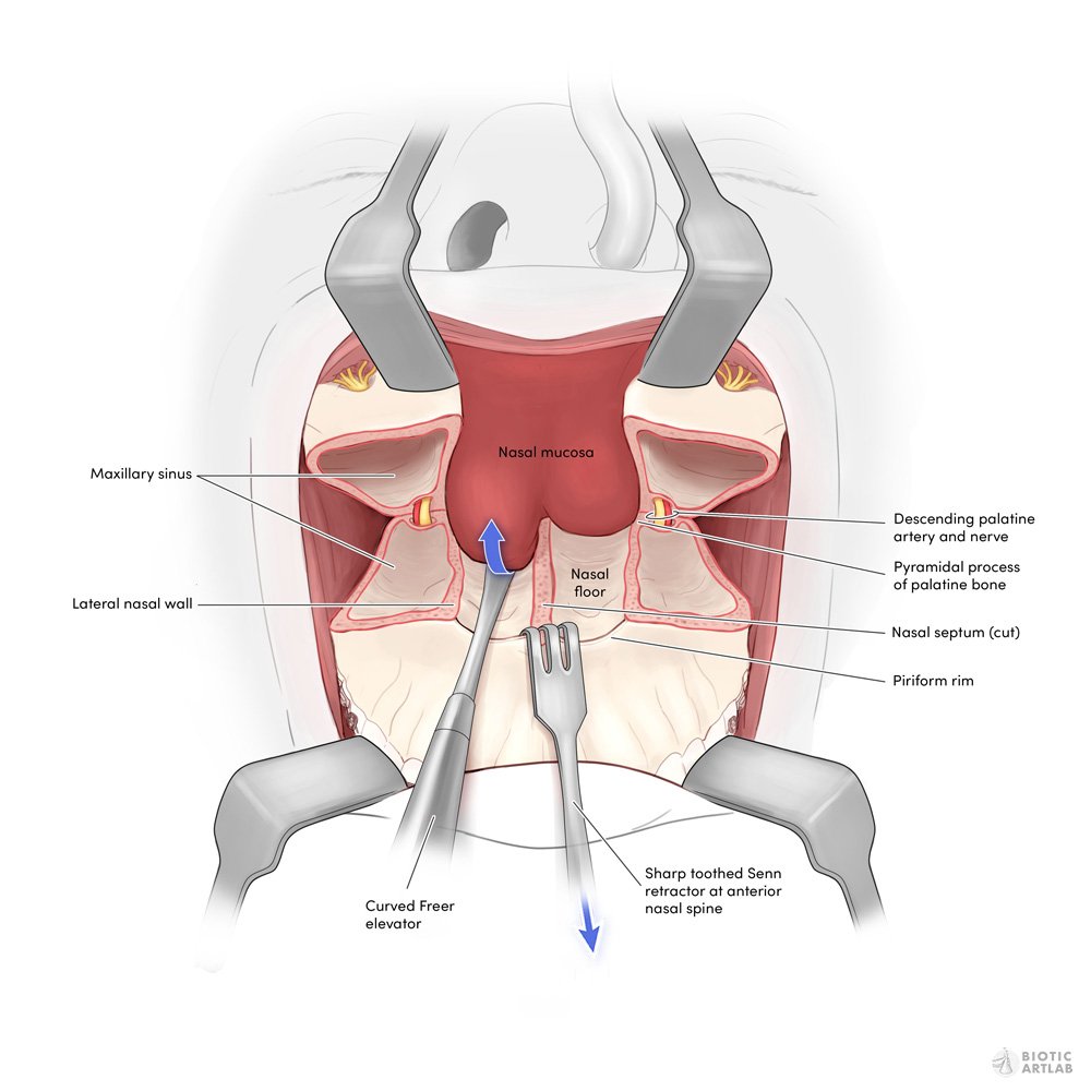
Step 4: Now there is access to the interior of the maxilla cavities.
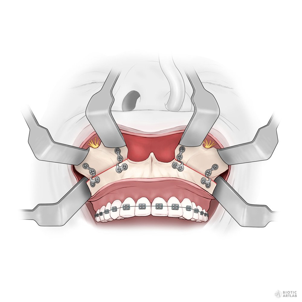
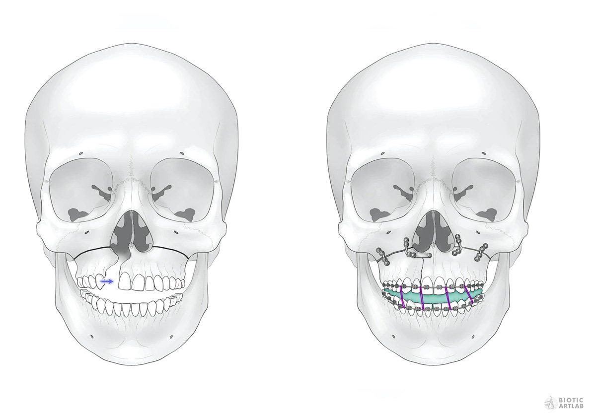
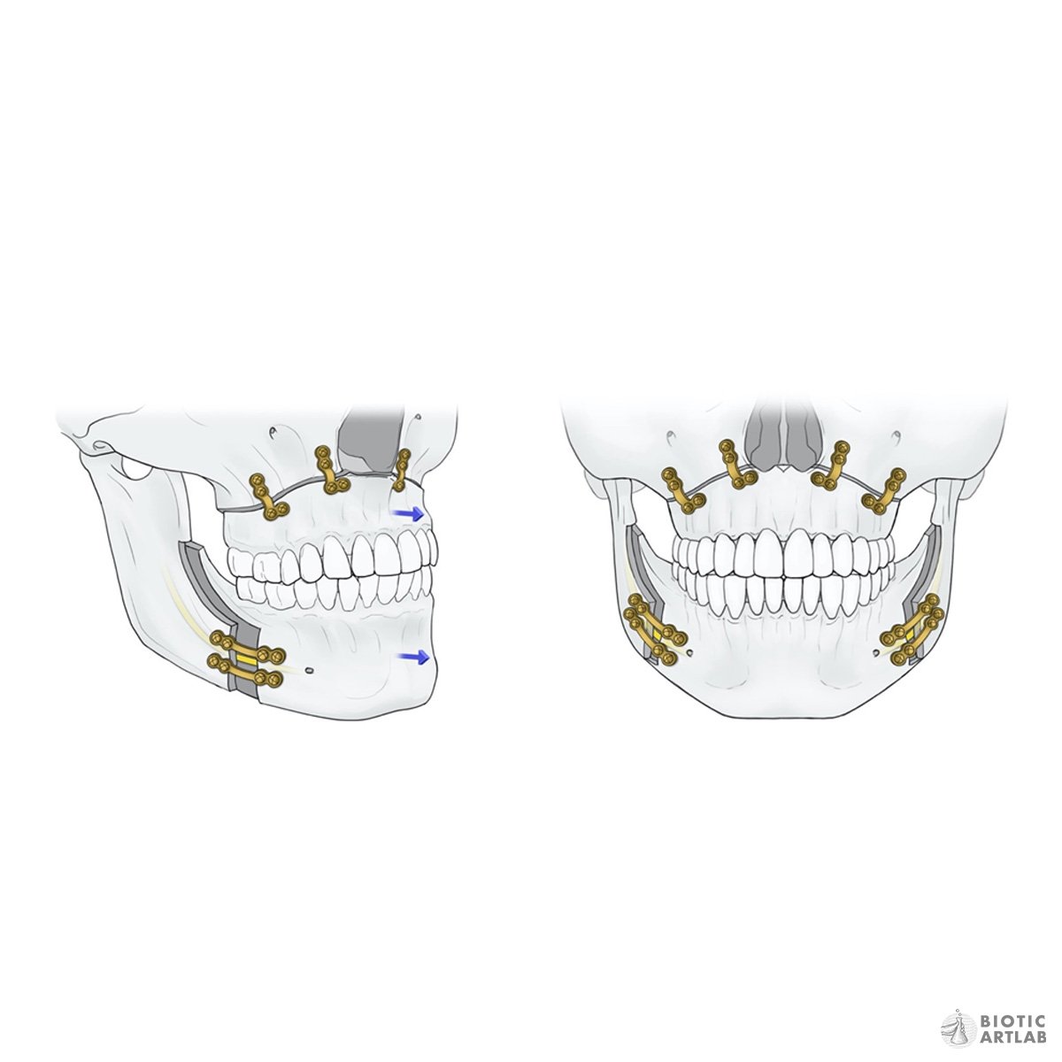
Step 5: Plates secure the maxilla in its new position.
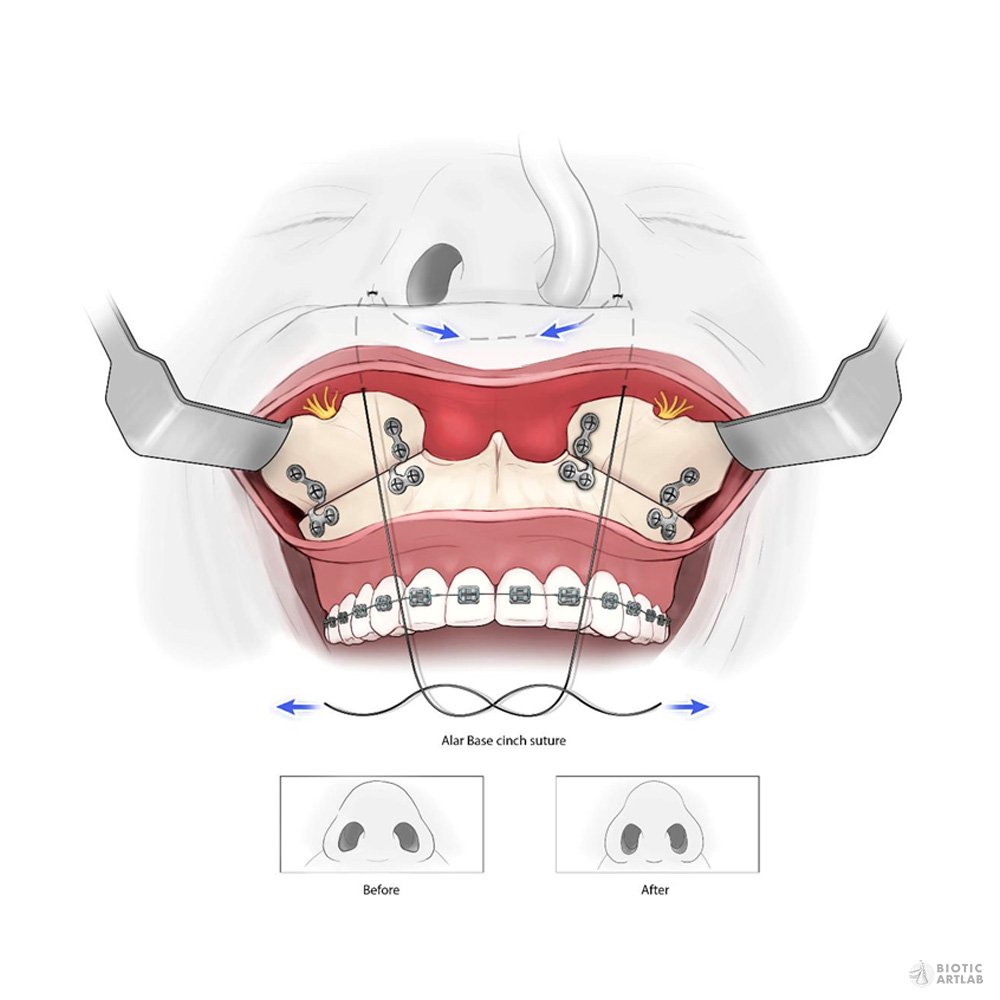
Step 6: An Alar Base cinch suture is used to re-narrow nose.
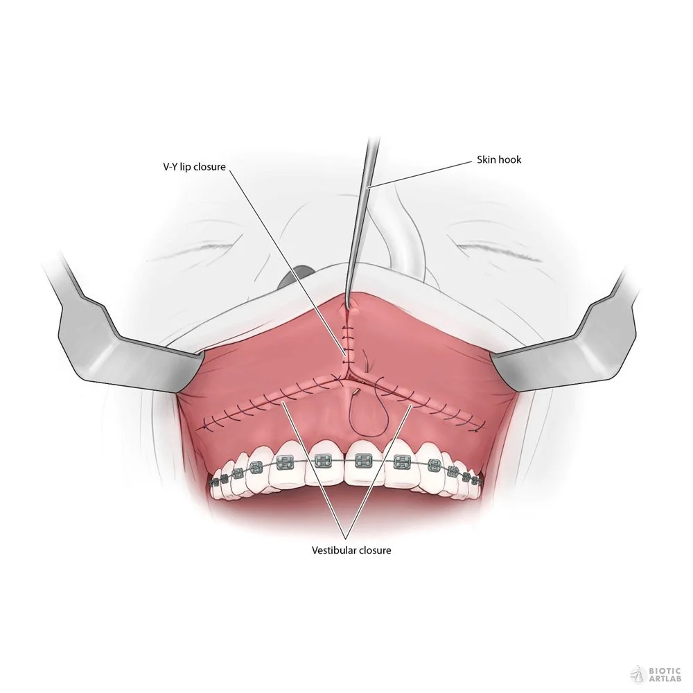
Step 7: V-Y suture used to close up soft tissue.
02 - What is a “maxillary osteotomy”?
When the bite of a patient’s teeth cannot be corrected by orthodontics alone, a maxillary osteotomy may be considered. This surgical procedure involves cutting through the bone of the upper jaw that then allows it to be repositioned with the lower jaw to close the bite. The jaw pieces are held in place with plates, screws, and sutures as it heals.

03 - Our Solution ✨
The incisions in the maxilla or mandible can vary greatly depending on the need of the patient’s bite. This required a deep dive into the history of maxillary osteotomies in order to understand how this surgical technique has evolved throughout time. To clearly convey the key elements of each procedure, we illustrated in greyscale and used accent colors to draw attention to important anatomical features - nerves to be aware of, teeth roots to avoid, and surgical cut lines to follow.
Let’s Chat
Are you looking to create educational material for your course ? Reach out to us and we can start the conversation.